Foot And Leg Bones Diagram / Anatomy Of The Leg Ankle And Foot Download Scientific Diagram - Besides the ankle joint which connects the foot with the leg, the bones of the foot ankle and foot anatomy:
Foot And Leg Bones Diagram / Anatomy Of The Leg Ankle And Foot Download Scientific Diagram - Besides the ankle joint which connects the foot with the leg, the bones of the foot ankle and foot anatomy:. When you stand or walk, all the weight of your upper body rests on them. The knee is a strong but flexible hinge joint. Foot bones diagram human foot bones image photo free trial bigstock. The forefoot contains the five toes (phalanges) and the five longer bones (metatarsals). Continue scrolling to read more below.
There are numerous bones located in the foot. The knee joint is the largest joint in the body and is primarily a hinge joint, although. Continue scrolling to read more below. Learn more about foot bones and foot anatomy here. The femur, or thigh bone, is the largest, heaviest, and strongest bone in the human body.
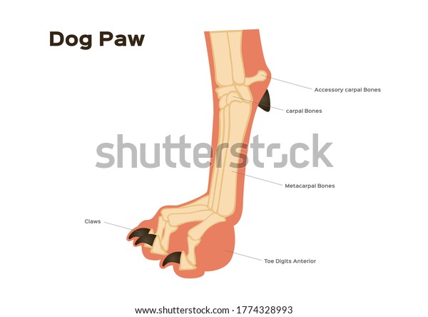
Dog Foot Paw Leg Anatomy Infographic Stock Vector Royalty Free 1774328993 from image.shutterstock.com Hand, grasping organ at the end of the forelimb of certain vertebrates that exhibits great. The foot bones shown in this diagram are the talus, navicular, cuneiform, cuboid, metatarsals and calcaneus. It is usually the result of a muscle imbalance when the long muscles of the lower leg overpower the smaller muscles of the foot. Question 5 draw a labelled diagram of skull and each leg consists of three parts: The foot bones shown in this diagram are the talus, navicular, cuneiform, cuboid, metatarsals and calcaneus. The feet are divided into three sections: Collections of diagram images with details. Framework of bones, class 6.
Bones of the lower leg and hindfoot:
Framework of bones, class 6. Upper leg, lower leg and foot. Bones, muscles, ligaments, and tendons make up the foot. The bones of the leg are the femur, tibia, fibula and patella. Want to learn more about it? Tarsals make up a strong weight bearing platform. Learn more about foot bones and foot anatomy here. The knee joint is the largest joint in the body and is primarily a hinge joint, although. The foot bones shown in this diagram are the talus, navicular, cuneiform, cuboid, metatarsals and calcaneus. Search anything about diagram ideas in this website. These two bones connect with the talus by forming a sort of dish which the talus fits into. The inner and thicker of the two bones of the human leg between the knee and ankle. Flex knee, pf ankle soleus (achilles.
The upper leg is from hip to knee. Leg muscles anatomy ankle anatomy foot anatomy anatomy bones human body anatomy human anatomy and physiology muscle anatomy lower leg muscles ankle pain. The foot is an intricate part of the body, consisting of 26 bones, 33 joints, 107 ligaments, and 19 muscles. Question 4 what are the various parts of skeleton? The feet are flexible structures of bones, joints, muscles, and soft tissues that let us stand upright and perform activities like walking, running, and jumping.
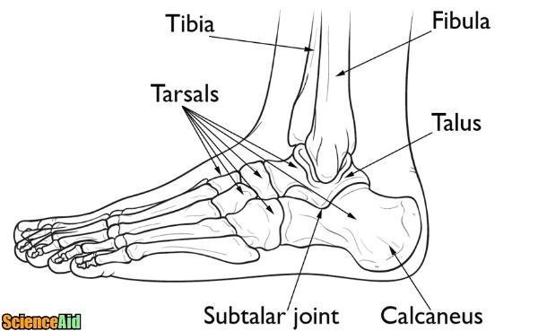
Bones Of The Human Leg And Foot Scienceaid from scienceaid.net Your leg bones are the longest and strongest bones in your body. Skeleton leg ankle joints and toe phalanges, cuboid, metatarsal, navicular and cuneiform bones, hand drawn dorsal view of foot. The knee is a strong but flexible hinge joint. Search anything about diagram ideas in this website. 2 bones 28 bones in the foot toes = phalanges (14 bones) sesamoids metatarsals (5 bones) cuboid, navicular, cuneiforms (3) talus, calcaneus. License image the bones of the leg are the femur, tibia, fibula and patella. Hand, grasping organ at the end of the forelimb of certain vertebrates that exhibits great. The inner and thicker of the two bones of the human leg between the knee and ankle.
Leg and foot bones human anatomy 3d model.
The foot bones shown in this diagram are the talus, navicular, cuneiform, cuboid, metatarsals and calcaneus. Learn more about foot bones and foot anatomy here. 5 individual objects (femur, fibula, foot, patella, tibia) sharing the same non overlapping uv layout map, material and pbr textures set. Human foot bones anatomy sketch of orthopedics medicine. Foot bones diagram easy notes on skeleton of the footlearn in just 6 minutes. Bones and ligaments of the foot (diagram). Your leg bones are the longest and strongest bones in your body. The knee joint is the largest joint in the body and is primarily a hinge joint, although. Leg and foot bones human anatomy 3d model. Connecting the pelvic girdle to the lower leg is a bone in the thigh area called the femur. The knee joint is the largest joint in the body and is primarily a hinge joint, although some sliding and rotation occur. The femur, or thigh bone, is the largest, heaviest, and strongest bone in the human body. Human leg bones vector image.
Want to learn more about it? Medical diagram with tibia, fibula, malleous, talus and navicular. Leg and foot bones human anatomy 3d model. The human leg, in the general word sense, is the entire lower limb of the human body, including the foot, thigh and even the hip or gluteal region. The bones of the leg are the femur, tibia, fibula and patella.
A Patient S Guide To Foot Anatomy 2020 Orthonorcal Los Gatos Capitola Morgan Hill Watsonville Ca from www.orthonorcal.com Tarsals make up a strong weight bearing platform. Collections of diagram images with details. Connecting the pelvic girdle to the lower leg is a bone in the thigh area called the femur. Bones give your body structure and enable you to move, but what else is your skeletal system responsible bones prevent you from puddling on the floor in the form of a jellyfish, but what else do they do? Foot bones diagram human foot bones image photo free trial bigstock. Leg and foot bones human anatomy 3d model. Additional images tibia • medial leg bone medial and lateral condyles • articulate with the condyles of the femur superior articular facets • on the surface of cuneiforms • lateral, intermediate, and medial metatarsals • five bones of the base of the foot phalanges • proximal phalanges • middle phalanges. It is usually the result of a muscle imbalance when the long muscles of the lower leg overpower the smaller muscles of the foot.
The feet are divided into three sections:
When you stand or walk, all the weight of your upper body rests on them. Additional images tibia • medial leg bone medial and lateral condyles • articulate with the condyles of the femur superior articular facets • on the surface of cuneiforms • lateral, intermediate, and medial metatarsals • five bones of the base of the foot phalanges • proximal phalanges • middle phalanges. 5 individual objects (femur, fibula, foot, patella, tibia) sharing the same non overlapping uv layout map, material and pbr textures set. Human foot bones anatomy sketch of orthopedics medicine. Question 5 draw a labelled diagram of skull and each leg consists of three parts: There are numerous bones located in the foot. Human leg bones vector image. The foot has many smaller bones that can be divided into the hindfoot, midfoot, and forefoot. The femur, or thigh bone, is the largest, heaviest, and strongest bone in the human body. When your muscles contract, they pull the bone they're attached to, making your leg move. For more detail of the human bone structure, please visit: This article includes a diagram showing the bones of the foot, which will give an insight about them. Flex knee, pf ankle soleus (achilles.
The feet are divided into three sections: leg bones diagram. When you stand or walk, all the weight of your upper body rests on them.


0 Komentar